2d echo normal result sample|Reference (normal) values for echocardiography : Pilipinas Two-dimensional (2D) ultrasound is the most commonly used modality in . The following list of postal codes for Tayabas, Philippines is derived from GeoNames.org. The data is provided "as is" without warranty or any representation of accuracy, timeliness or completeness. 4327 Tayabas. Google Map for Tayabas, Philippines GPS coordinates: 14.0289, 121.5911.For many, when you mention New York, the immediate image that springs to mind is the bustling metropolis of New York City – the city that never sleeps. However, New York is a vast and diverse state, boasting more than just one iconic region. Today, we’re exploring a less globally renowned, but equally captivating corner of the state: the .

2d echo normal result sample,Normal values for aorta in 2D echocardiography. Normal interval. Normal interval, adjusted. Aortic annulus. 20-31 mm. 12-14 mm/m2. Sinus valsalva. 29-45 mm. 15-20 .Two-dimensional (2D) ultrasound is the most commonly used modality in .Usually normal (LVEDV ≤150ml male, ≤106ml female) Normal or mild dilated: Dilated 2 (LVEDV >150ml male, >106ml female) Left atrium (Size) 1: Usually normal (LA volume ≤34ml/m 2) Normal or mild dilated: Dilated .
Below is a complete and thorough list of normal echo values. This list of normal echo values is from echopedia.org. Left Ventricle. Left Ventricular Systolic Function. Left Ventricular Diastolic .measurement of aortic root dimensions.1,2 Normal reference ranges have been mainly established for two-dimensional (2D) echocardiography with fundamental imaging .
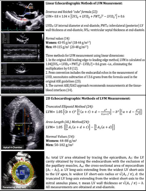
A normal EF is about 55-65 per cent. It’s important to understand that “normal” is not 100 per cent. Measuring the EF helps your doctor to understand how well the heart is .Reference (normal) values for echocardiography2D methods: truncated ellipsoid method or area length method. 50-102: 103-116: 117-130 > 130: Relative wall thickness (cm)* = 2 x posterior wall / left ventricular diastolic diameter. 0,24-0,42: 0,43-0,46: 0,47-0,51 > 0,51
With regards 3D volumetric assessment and 3D-derived ejection fraction, the BSE stresses that reference intervals for 2D-derived ejection fraction do not apply to .
Frequently Asked Questions: What is the 2D Echo Test used for? 2D Echo test is a non-invasive diagnostic test used for detecting various cardiac abnormalities like. A defect in . A normal test result reflects normal functioning; structure; and movement of heart muscles, valves and chambers. An abnormal 2D ECHO (both TTE and TEE) test could indicate a myriad of heart problems. It requires consultation with a cardiologist for a final diagnosis and treatment. A normal stress echocardiogram reveals that the heart .
An echocardiogram (or echo) is an ultrasound of the heart. During an echo, we record short videos of the heart as it beats, and from these videos we can learn about the structure and function of the heart. The left ventricle is the main pumping chamber of your heart – it is the one where blood leaves your heart to be pumped around your body. Transthoracic echocardiography (TTE), sometimes called “surface echocardiography,” is a basic tool for investigation and follow-up of heart disease. Consultants who interpret TTE endeavour to provide accurate, useful reports to colleagues who order these tests. Referring physicians sometimes find reported results difficult to .
2 likes. A 2D echo test, also known as two-dimensional echocardiography, is a non-invasive procedure that utilises ultrasound waves to evaluate the function and structure of your heart. The images generated by this procedure are referred to as echocardiograms. Through this test, doctors can observe the real-time motion of the heart and assess . Two-dimensional (2D) or three-dimensional (3D) echocardiogram. These images provide pictures of the heart walls and valves and of the large vessels connected to your heart. A standard echocardiogram begins with a 2D study of the heart. A 3D echocardiogram is available in some medical centers and hospitals. It's often done to . The 2D Echo. An echocardiogram, or 2D echo or heart ultrasound an ultrasound examination that uses very high frequency sound waves to make real time pictures and video of your heart. Things that will be seen during a 2D echo test are the heart’s chambers, heart valves, walls and large blood vessels that are attached to your .
Two-dimensional (2D) or three-dimensional (3D) echocardiogram. A 2D echo is the standard test, which shows your doctor images of your heart's walls, valves, and some vessels. Results Demographic data. Table 1 summarizes the demographic data obtained in the entire population and according to gender. By inclusion criteria, patients had normal anthropometric and biological characteristics. A total of 198 men (44.1%) (mean age: 45.9 ± 14.0 years) and 251 women (55.9%) were included (mean age: 45.7 ± 13.4 .
Two-dimensional echocardiography (2D echo) is a non-invasive diagnostic technique that provides information regarding the cardiac function and hemodynamics. . The practice of clinical .

Information from an echocardiogram may show: Changes in heart size. Weakened or damaged heart valves, high blood pressure or other diseases can cause thickened heart walls or enlarged heart chambers. Pumping strength. An echocardiogram can show how much blood pumps out of a filled heart chamber with each heartbeat.901 West 43rd St. Telephone: 816-569-2200 Kansas City, MO 64111 www.sononet.us Fax: 816-581-2090.

Echocardiography is the use of ultrasound to evaluate the structural components of the heart in a minimally invasive strategy. Although, prior to the invention of today's routinely used 2-dimensional .
The results of an echocardiogram are responsible to project the actual real-time images of a heart, if its functionality is normal or abnormal, its pumping strength, etc. The walls of a heart is one of the things that can be seen very clearly during an echocardiogram – according to experts if these walls are measured anything thicker .2d echo normal result sample An echocardiogram—echo for short—is an ultrasound of your heart. Echocardiograms show the size and structure of the heart and what’s happening in the different chambers as your heart is beating. Keep in mind that an echo is one method a cardiologist uses to make a diagnosis. Your cardiologist will interpret the results in the .
An echocardiogram is a specialized ultrasound scan of the heart. It gives detailed information about how efficiently the heart pumps blood and oxygen to the organs and how well the heart valves work. A trained and qualified cardiologist (heart specialist) needs to read an echocardiogram to know if the results are normal or not.Echocardiogram. An echocardiogram, or "echo", is a scan used to look at the heart and nearby blood vessels. It's a type of ultrasound scan, which means a small probe is used to send out high-frequency sound waves that create echoes when they bounce off different parts of the body. These echoes are picked up by the probe and turned into a moving .An echocardiogram (echo) is a test that diagnoses and manages heart disease. An echo uses ultrasound to create pictures of your heart’s valves and chambers. 800.223.2273; . For example, some people with valve disease need echo tests on a regular basis.
Normal values and thresholds for all heart structures including illustrations . Echocardiography in aortic diseases: EAE recommendations for clinical practice. In European journal of echocardiography : the journal of the Working Group on Echocardiography of the European Society of Cardiology 11 (8), pp. 645–658. . (g/m²) .
2d echo normal result sample|Reference (normal) values for echocardiography
PH0 · What is a 2D Echo Test?
PH1 · What is 2d Echo test and its Uses, Test Results, and Normal
PH2 · Two
PH3 · Reference (normal) values for echocardiography
PH4 · Normal reference intervals for cardiac dimensions and function
PH5 · Normal Echo Values
PH6 · How to read an echocardiogram report
PH7 · How To Read The 2D Echo Test Result? What Does It Show
PH8 · How To Read The 2D Echo Test Result? What Does It Show
PH9 · Echocardiography (Normal values)
PH10 · Echocardia